黄石免疫电镜英文
- tem电镜样品
- 2024-05-11 00:00:28
- 224
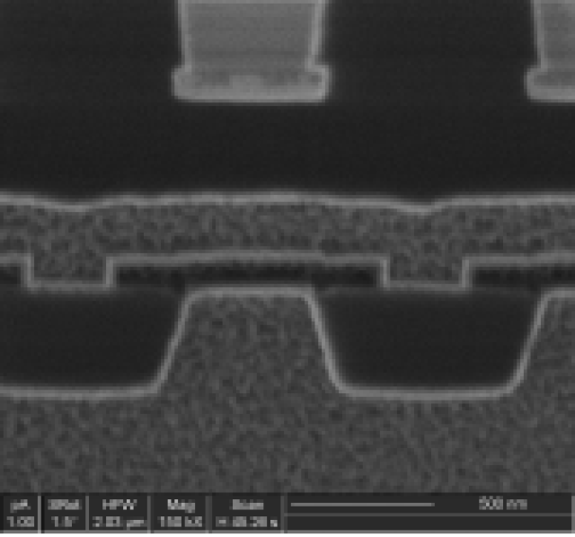
纳瑞科技的服务将为IC芯片设计工程师、IC制造工程师缩短设计、制造时间,增加产品成品率。我们将为研究人员提供截面分析,二次电子像,以及透射电镜样品制备。我们同时还为聚焦离子束系统的应用客户提供维修、系统安装、技术升级换代、系统耗材,以及应用开发和培训。
Title:Immunofluorescence and Electron Microscopy: A Powerful Combination for Studying Cellular Structures

Immunofluorescence and electron microscopy are two widely used techniques in modern cell biology. While immunofluorescence is primarily used to visualize specific proteins within cells, electron microscopy is used to capture high-resolution images of cell structures. The combination of these two techniques offers researchers a unique opportunity to explore cellular structures at the molecular level. In this article, we will discuss the principles of immunofluorescence and electron microscopy, their applications, and the ways in which they can be combined to gain a comprehensive understanding of cellular structures.
I. Introduction
Immunofluorescence and electron microscopy are two powerful tools that enable researchers to study the intricate details of cellular structures. Immunofluorescence is a technique that combines the ability of antibodies to recognize specific proteins with the sensitivity of in situ hybridization, allowing for the visualization of protein-rich structures within cells. Electron microscopy, on the other hand, provides high-resolution images of cellular structures at the electron microscopic level, enabling researchers to capture detailed images of cell membranes, organelles, and other structures.
By combining immunofluorescence and electron microscopy, researchers can gain a comprehensive understanding of cellular structures at the molecular level. This technique, often referred to as immunoelectron microscopy, offers researchers a unique opportunity to explore the complex dynamics of cellular structures in real-time.
II. Immunofluorescence
Immunofluorescence is a technique that combines the use of antibodies and in situ hybridization with electron microscopy. In this technique, antibodies are first introduced into cells, either through注射 or through the use of transfection. These antibodies bind specifically to specific proteins, either by their primary or secondary structure. These bound proteins can then be visualized using electron microscopy, allowing researchers to capture detailed images of the structures.
One of the key advantages of immunofluorescence is its ability to provide high-resolution images of specific proteins within cells. This technique is particularly useful for studying the organization of cell structures, such as cell membranes, organelles, and the cytoskeleton. By visualizing specific proteins within these structures, researchers can gain insights into the mechanisms that govern their dynamics and functions.
III. Electron Microscopy
Electron microscopy is a technique that uses a beam of electrons to produce high-resolution images of cellular structures. In this technique, a sample is placed in a vacuum, and a beam of electrons is directed onto the sample. The electrons interact with the sample, resulting in the emission of electrons, which are then detected by an image plate. This interaction provides high-resolution images of the structures within the sample.
One of the key advantages of electron microscopy is its ability to capture detailed images of cellular structures at the electron microscopic level. This technique is particularly useful for studying the organization of cell membranes, organelles, and the cytoskeleton. By visualizing these structures at the molecular level, researchers can gain insights into their functions and the mechanisms that govern their organization.
IV. Combining Immunofluorescence and Electron Microscopy
Immunofluorescence and electron microscopy can be combined in various ways to enable researchers to explore cellular structures at the molecular level. One common method is to use immunofluorescence to visualize specific proteins within cells, and then use electron microscopy to capture detailed images of the structures. This combination allows researchers to gain a comprehensive understanding of cellular structures and their organization.
Another method is to use electron microscopy to capture images of cellular structures, and then use immunofluence to visualize specific proteins within those structures. This approach enables researchers to gain insights into the mechanisms that govern the organization of cellular structures, as well as the functions of the proteins within those structures.
V. Conclusion
Immunofluorescence and electron microscopy are two powerful tools that enable researchers to study cellular structures at the molecular level. By combining these techniques, researchers can gain a comprehensive understanding of cellular structures and their organization. This combination is particularly useful for studying the organization and dynamics of cell membranes, organelles, and the cytoskeleton.
In the future, immunoelectron microscopy will likely become a standard tool for studying cellular structures. The ability to visualize specific proteins within cells and capture detailed images of cellular structures at the electron microscopic level will enable researchers to gain insights into the mechanisms that govern cellular structures and their organization.
Combining immunofluorescence and electron microscopy is a powerful tool for studying cellular structures. By gaining a comprehensive understanding of cellular structures at the molecular level, researchers can gain insights into the mechanisms that govern their organization and functions. Further research